I often meet patients who arrive at my clinic looking rather worried, clutching an MRI report filled with alarming medical terminology.
"Doctor, I'm only 30, but apparently I have the spine of a 90-year-old!"
I've heard variations of this more times than I can remember.
The report might mention degenerative discs, disc bulges, arthritis, narrowing of the spinal canal and perhaps a few other rather unpleasant-sounding findings.
And after spending an evening consulting Dr Google, things have usually become considerably worse! (And yes, I know I'm taking a cheeky little swipe at Dr Google here.)
But here's something I often explain to my patients: an MRI tells us only part of the story.
What does an MRI actually show?
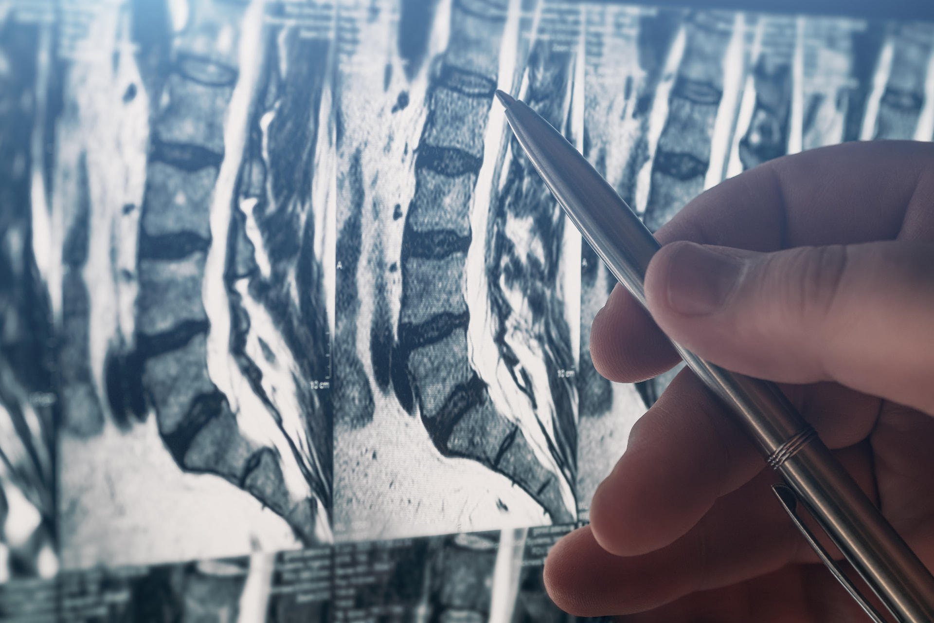
An MRI is a remarkable piece of technology. It allows us to see the structures inside our bodies in extraordinary detail, including discs, nerves, muscles and joints.
It is particularly useful when we are looking for significant abnormalities, such as nerve compression, spinal narrowing, infection, tumours or other conditions that might require specific treatment.
But an MRI is essentially a photograph of your anatomy.
It cannot tell us exactly how much pain you experience, how well you sleep, whether you can comfortably walk around the supermarket or how your symptoms affect your everyday life.
And perhaps most importantly, not everything that appears abnormal on an MRI is necessarily responsible for your pain.
Does everyone have a disc bulge?
Well, not quite everyone. But you might be surprised!
Research has shown that disc degeneration, bulging discs and other spinal changes are remarkably common, even in people who have absolutely no back pain.
In one large review, approximately 40% of people in their thirties without back pain had a disc bulge on imaging.
Yes, you read that correctly. They had these findings without experiencing any symptoms!
Our bodies change as we grow older. Just as our hair turns grey and our skin develops wrinkles, our spines undergo structural changes over time.
A disc bulge does not automatically mean your spine is damaged beyond repair, nor does the word "degeneration" mean your back is falling apart.
Of course, certain findings can be clinically important. The challenge is determining which ones actually matter.
The 10% and 90% rule
I often tell patients that imaging gives me perhaps 10% of the story, while the remaining 90% comes from the clinical assessment.
Now, these aren't scientifically calculated percentages! It's simply my way of explaining how important the clinical history and examination are.
I want to know where your pain is, how it started, what makes it worse and what helps. I examine how you move, assess your nerves and try to understand how the pain affects your life.
Only then do I interpret the imaging in that context.
For example, a disc pressing on a nerve may be highly relevant when someone has matching leg pain, weakness and neurological findings.
The same scan finding in someone without those symptoms may be far less important.
This doesn't mean I disregard imaging. Quite the opposite. I use it to help answer specific clinical questions rather than allowing the scan to dictate the entire diagnosis.
But my MRI is normal, and I'm still in pain!
Now, let's consider the opposite situation.
Imagine living with severe back pain for months. You've struggled to work, sleep or enjoy your usual activities. Eventually, you undergo an MRI, hoping it will finally explain everything.
And the report comes back as essentially normal. Yes, plum normal!
Rather than feeling reassured, you might actually feel disappointed. After months of pain, you were hoping the scan would finally give you an answer. Instead, you're left wondering why you're hurting so much when everything apparently looks fine.
If nothing is wrong on the scan, why does it hurt so much?
Pain is a complex experience involving the nervous system, tissues and the way our brain processes incoming signals.
Not every source of pain is visible on imaging. Changes in nerve sensitivity, muscle function and pain processing may contribute to symptoms without producing a clear structural abnormality.
A normal MRI is reassuring in many respects, but it does not mean your pain is imaginary, exaggerated or made up.
Nor should it automatically lead us to conclude that the problem is psychological.
It simply tells us that the particular investigation has not identified a structural explanation for your symptoms.
So, do I actually need an MRI for every chronic pain issue?
Not necessarily.
Many episodes of pain can be assessed and managed without advanced imaging, particularly when there are no concerning clinical features.
I usually consider imaging when it may help identify significant pathology, clarify a diagnosis or guide an important treatment decision.
There are circumstances where imaging becomes particularly important, including progressive neurological weakness, suspected infection, fracture or malignancy. New bladder or bowel dysfunction with concerning neurological symptoms requires urgent medical assessment.
The decision should be guided by your symptoms and examination, rather than simply how long you've been in pain.
Looking beyond the scan
I believe good pain medicine begins with listening to the patient, not just reading the radiology report.
An MRI can provide valuable information, but it should complement our clinical assessment rather than replace it.
An alarming-looking scan does not necessarily mean your future is bleak. Equally, a reassuring scan does not invalidate the pain you are experiencing.
Ultimately, I treat the person sitting in front of me, not the scan report.
And fortunately, your spine doesn't come with an expiry date!
Best wishes

This article is intended for general information and education only. It is not a substitute for individual medical advice, diagnosis or treatment. The appropriateness of any investigation, medication, supplement, procedure or other treatment depends on your individual circumstances and should be discussed with your treating healthcare professional.